


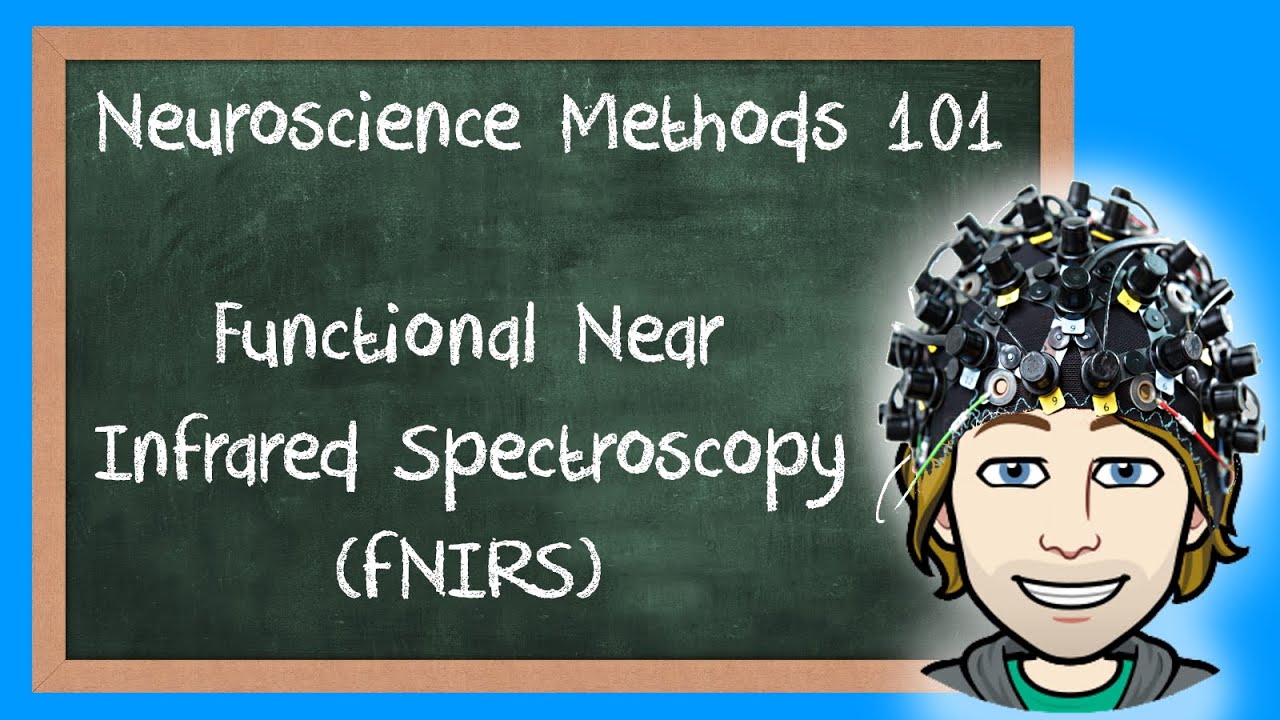
Functional Near Infrared Spectroscopy (fNIRS) Explained! | Neuroscience Methods 101
101 today were going to talk about functional, near infrared, spectroscopy or fners. In neuroscience, we often want to know which brain regions are activated during a specific action. Previously, we have seen how functional magnetic resonance, imaging or fmri can give you colorful pictures of the brain. The colors actually represent regions in the brain, with increased or decreased oxygen levels and since neurons in the brain need oxygen to work, fmri basically tells you at what locations neurons are active. So if we already have a method fmri, why do we need something else? Well, fmri is very expensive, but maybe, more importantly, in an mri scanner. You have to lie down and you are not allowed to move. This restriction can be problematic in certain studies, for example, when working with patients or with children, for whom it is very difficult to lie still for an hour. Also, some research questions can only be addressed if there is some movement. For example, when there is a movement learning task for these situations, there is fniers just as fmri fniers measures the oxygen levels in the brain, specifically the so called blood, oxygenation level, dependent response or, in short, the bold response, just after our neurons were active. They need to be replenished with energy and thus oxygen, so the bold response is a sharp rise in oxygen levels in a region of the brain that was active a few seconds earlier. But how does fneers pick up these oxygen levels for that? It uses infrared light.
Specifically near infrared light, whereas visible light barely penetrates the body infrared light can travel through the skin and bone, particularly near infrared waves, which have wavelengths that are closest to visible red light near infrared waves, penetrate the body and reach the brain. But not everything in the body lets near infrared light through equally, some mediums absorb the waves at different, specific wavelengths. The cool thing is that oxygen in the blood absorbs infrared light as well, so blood with a lot of oxygen or oxygenated blood absorbs light with longer wavelengths above 790 nanometers, but blood without oxygen or deoxygenated. Blood absorbs slight with shorter wavelengths wavelengths below 790 nanometers fneers uses exactly these physiological features, so an f nearest machine has light emitting diodes that send the light source through the head and the skull. Additionally, the afners has light detectors that detect how much of the light at a certain wavelength is absorbed. The leds use multiple lights at different wavelengths within the near infrared range. Some of them have longer near infrared wavelengths and thus should be absorbed by oxygenated blood and others have shorter near infrared wavelengths, which should be more absorbed by deoxygenated blood. After the light detectors pick up the absorption rate at different wavelengths, a ratio of oxygenated and deoxygenated blood can give an indication of which brain areas were active at a specific time point. So , both fniers and fmri measure the blood oxygenation level dependent response, but they do it in a different way and, as mentioned before, fmri is very expensive, whereas f nearest is quite low in cost.
But since fniers depends on the select number of electrodes. Fmri is much more precise. The spatial resolution of fmri is in the range of a couple of millimeters, whereas the spatial resolution of fniers is more in the range of a couple centimeters. Having said that, f nearest is less sensitive to motion and thus it is ideal for pediatric research. So, in the end, if f neers is the right method will depend on the research question, so i hope you had fun.


